A "perfect storm" of an increased emphasis on patient-centered care, decreasing reimbursements and improving technology has set the stage for radiologists to make MRI more efficient.
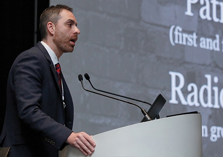
This helps explain the evolution of fast MRI, according to Soterios Gyftopoulos, MD, associate professor in musculoskeletal radiology and orthopedic surgery at NYU Langone Medical Center. He moderated a Wednesday "Hot Topics" session devoted to fast musculoskeletal MR imaging.

Gyftopoulos
The emphasis on patient-centered care comes into play, Dr. Gyftopoulos said, with the fact that fast MRI provides a better experience for the patients. "Most people don't want to be in an MR scanner for 30 minutes," he said. "It's an uncomfortable location. It's noisy and you can't move."
Instead, fast MRI dramatically decreases the amount of time it takes to perform an exam.
"The patient only has to stay still for 4 or 5 minutes, and radiologists still get the same amount of imaging information they need to make an accurate diagnosis," Dr. Gyftopoulos said.
Patient Comfort Helps Image Quality
It can also improve image quality, he said, particularly in cases where radiologic technologists are dealing with difficult patients.
For example, in the past the technologists might have simply powered through the exam and completed the study with the help of the patient, but, possibly at the cost of image quality.
"Fast MRI can help with this by being a problem solver," he said, explaining that technologists now know to run a fast protocol in these cases. "This has resulted in better quality exams for these patients."
And in a time when imaging centers and departments are under increasing pressure due to reimbursement cuts, fast MRI provides them with a way to become more efficient, perform more exams and increase revenue, Dr. Gyftopoulos said. "Which, in turn, allows us to invest in better equipment and facilities, in our resident and fellowship programs, and in research that can change the field of imaging."
Speaking on emerging techniques for fast MR imaging, Jan Fritz, MD, associate professor of radiology and radiologic science, Johns Hopkins University, noted that one of the workhorses in current 2D MR imaging practice is parallel imaging acceleration, which is excellent for speed, but pays a penalty in signal-to-noise, he said.
One promising technique, he said, is to combine parallel imaging with simultaneous multi-slice (SMS) acquisition. In a study published last year in Investigative Radiology, Dr. Fritz and his colleagues found that four-fold-accelerated, high-resolution turbo spin echo MRI of the knee, through the combination of parallel imaging and SMS, resulted in 50 percent shorter acquisition times compared to regular parallel imaging acceleration, with similar image quality.
"This is a great way to appropriately use our imaging time," Dr. Fritz said. "I think it's the way to go if you are a fan of 2D imaging."

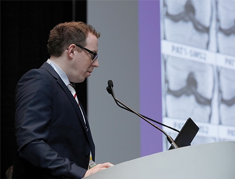
Fritz
How Fast Can MRI Go?
Dr. Fritz compared three MR images of a meniscus tear — one 2D image acquired in slightly less than 4 minutes, a 3D image acquired in four minutes, 46 seconds, and a 3D image acquired in 90 seconds with compressed sensing.
There was visible degradation in the 90-second image, Dr. Fritz said, but, arguably, you can still see the posterior meniscus tear, and you can do an isotropic data set with 0.5 isotropic resolution in 90 seconds.
"I think this is where we are going," he said. "At some point MR imaging is going to get incredibly fast."

